Metagenomics Metadata and Metaanalysis


I am a co-organizer of this… see for yourself. Cutting edge metagenomics research, discussion of standards applications to genomics and metagenomics, all in beautiful Stockholm this summer. If you are coming to ISMB/ECCB 2009, consider coming to the M3 SIG.

Link to announcement 1-page poster, because you really want to tack this to your departmental bulletin board: m3-poster

m3_icon_main

Call For Abstracts and Posters:  Metagenomics, Metadata and Metaanalysis

An ISMB / ECCB 2009 SIG

Date: June 27, 2009

Location: Stockholm, Sweden

URL: http://gensc.org/gc_wiki/index.php/M3

There are now thousands of genomes and metagenomes available for study. Interest in improved sampling of diverse environments (e.g. ocean, soil, sediment, and a range of hosts) combined with advances in the development and application of ultra-high throughput sequence methodologies are set to vastly accelerate the pace at which new metagenomes are generated.

Continue reading Metagenomics Metadata and Metaanalysis →

Share and Enjoy:
  • Fark
  • Digg
  • Technorati
  • del.icio.us
  • StumbleUpon
  • Facebook
  • Reddit
  • Twitter
  • FriendFeed
  • PDF
  • email
  • Print
  • Google Bookmarks

Naturally Obsessed

A new documentary film follows life in a molecular biology lab in Columbia University over the course of three years. It looks very promising: the title is certainly something many of us identify with.

Share and Enjoy:
  • Fark
  • Digg
  • Technorati
  • del.icio.us
  • StumbleUpon
  • Facebook
  • Reddit
  • Twitter
  • FriendFeed
  • PDF
  • email
  • Print
  • Google Bookmarks

Now this looks a lot better!


ResearchBlogging.org
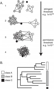
A network representation  (A) vs. a tree representation (B) of protein sequence similarities. Click for larger picture & caption.

A network representation (A) vs. a tree representation (B) of protein sequence similarities. Click for larger picture & caption. Source: doi:10.1371/journal.pone.0004345

In any data-rich science, data visualization is of prime importance.  Finding ways to visually depict data is challenging, as we have opposing demands: we would like to see the data in the whole, but also be able to zoom in and analyze the details; we would like to know how the many details add up to affect the big picture.

There are already templated ways of presenting  molecular biology data. Gene expression data is typically shown in clustered heat maps; phylogenetic data is shown in trees (one of the oldest ways of representing data rich sets in a graphic manner — dating back to Linnaeus in the mid 18th century, although he used it for taxonomy), and sequence similarities are shown in color-coded multiple sequence alignments and as trees. Protein-protein interaction data is shown as graphs with the nodes being individual proteins and vertices drawn between interacting proteins.

But sometimes, borrowing method A to represent data of type B has unusual benefits.

Continue reading Now this looks a lot better! →

Share and Enjoy:
  • Fark
  • Digg
  • Technorati
  • del.icio.us
  • StumbleUpon
  • Facebook
  • Reddit
  • Twitter
  • FriendFeed
  • PDF
  • email
  • Print
  • Google Bookmarks

Music: Vic Ruggiero and Lisa Mueller

These two have great chemistry, and the video captures the raw and gritty quality of a small club appearance.

Share and Enjoy:
  • Fark
  • Digg
  • Technorati
  • del.icio.us
  • StumbleUpon
  • Facebook
  • Reddit
  • Twitter
  • FriendFeed
  • PDF
  • email
  • Print
  • Google Bookmarks

Enzyme Promiscuity

ResearchBlogging.org
Gradualism is a cornerstone principle in evolution.  Things happen in small increments;  all the time that changes happen, overall organism fitness cannot be compromised.  So how then do full-featured complex functions appear? One way is by functional re-purposing of an existing organ:
…an organ may become rudimentary for its proper purpose, and be used for a distinct object: in certain fish the swim-bladder seems to be rudimentary for its proper function of giving buoyancy, but has become converted into a nascent breathing organ or lung. Other similar instances could be given.
— Charles Darwin Origin Ch. 13
(Actually, today experts think it’s the other way around: that the swim bladder evolved from the lungs of bony fish… but the principle still applies).
A similar re-purposing phenomenon happens in enzymes, and the mechanism by which it appears was dubbed enzyme promiscuity. The idea behind enzyme promiscuity is simple: an enzyme E may catalyze a reaction S1 –>P1
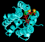
<em>C. freundii</em> DHAK structure. Cyan: ATP binding domain. Red: ATP. Green: ATP binding residues on the protein. Orange speres: Mg<sup>2+</sup> atoms

C. freundii DHAK structure. Cyan: ATP binding domain. Red: ATP. Green: ATP binding residues on the protein. Orange spheres: Mg2+ atoms. Click for larger image.

(substrate  1 to product 1) with a certain degree of efficiency. However, E  may also  catalyze S2 –> P2 albeit with much lesser efficiency.  Now suppose the gene for this  enzyme E gets duplicated in the genome, not an uncommon occurrence: one gene copy E1 goes ahead through the generations catalyzing the original reaction S1–>P1. The second copy, E2, is now superfluous  for performing S1–>P1. E2 is now “free to evolve”, with no fitness constraints tying it to the original function.  If  the new reaction S2–>P2 increases the organism’s fitness, then E2 is under positive selection, for mutations increasing its ability to create P2 from S2.  For example, P2 may be a rare nutrient, but S2 a plentiful one, thus any organism that can make its own P2 is at an advantage in S2-poor environments.
In a  very elegant work published this week in Chembiochem,  Eduardo Jucenda and his colleagues have captured a snapshot of the evolution of enzyme promiscuity, with the old function maintained, the new one evolving, and  without gene duplication necessary.  The”E1″ discussed is dihydroxyacetone kinase or DHAK, which the group has been researching in the bacterium C. freundii. Like many enzymes, DHAK uses a metal ion as a cofactor to perform its catalysis, in this case magnesium (Mg2+).  During that time another group, Cameselle and co-workers have reported that another enzyme, FMN cyclase isolated from humans, is functionally promiscuous: it catalyzes both a DHAK reaction and an FMN cyclase reaction. FMN cyclase uses manganese ion (Mn2+) as a cofactor. Since the sequence of the human FMN cyclase has a 40% sequence identity with their bacterial DHAK, Jucenda’s group have decided to investigate if the converse is true: whether the bacterial DHAK can catalyze an FMN-cyclase reaction.  They discovered that the bacterial DHAK can perform FMN-cyclase activity, but it needed Mn2+ as a cofactor (remember: bacterial DHAK uses Mg2+).  Furthermore,when going above a certain Mn2+ concentration, the kinase activity is inhibited, while the cyclase activity is not (although the cyclase activity in the bacterial enzyme is much weaker than in the rat enzyme). So what Jucenda’s group has discovered  was not just a promiscuity event, but a way to switch between two different activities by changing the concentration of Mn2+ in the reaction. This switch promotes activity 2 (cyclase) while maintaining activity 1 (DHAK), in the absence of gene duplication, this mechanism can maintain both activities. C. Freundii can actually raise it’s Mn2+ concentration temporarily, which supports the hypothesis that the researchers have discovered a new mechanism for switching between cyclase and kinase activities, and its mechanism of evolution.

Further reading:

Israel Sánchez-Moreno, Laura Iturrate, Rocio Martín-Hoyos, María Luisa Jimeno, Montaña Mena, Agatha Bastida, Eduardo García-Junceda (2009). From Kinase to Cyclase: An Unusual Example of Catalytic Promiscuity Modulated by Metal Switching ChemBioChem, 10 (2), 225-229 DOI: 10.1002/cbic.200800573

A very interesting review on enzyme promiscuity:

O KHERSONSKY, C ROODVELDT, D TAWFIK (2006). Enzyme promiscuity: evolutionary and mechanistic aspects Current Opinion in Chemical Biology, 10 (5), 498-508 DOI: 10.1016/j.cbpa.2006.08.011

Share and Enjoy:
  • Fark
  • Digg
  • Technorati
  • del.icio.us
  • StumbleUpon
  • Facebook
  • Reddit
  • Twitter
  • FriendFeed
  • PDF
  • email
  • Print
  • Google Bookmarks

Are you stupid enough to work here?

Almost every time you read an interview with a scientist, the subject turns invariably to their sense of accomplishment and awe when they finally discover something new. Somehow, I never could quite identify with this sentiment entirely. Research is excruciatingly hard, laborious, frustrating and accomplishing anything always takes longer than I thought. I always keep hitting blind alleys; things don’t work and I don’t know why. I work for a long time on trying to confirm an hypothesis only to realize that I took a wrong turn near Albuquerque and I wasted time and money on what in 50/50 hindsight is an obviously stupid decision (and it seemed such a great idea two months ago!). By the time my findings are actually valid, novel and confirmed, i.e. “publishable”, whatever awe and wonder exists at this final stage is heavily tainted by exhaustion from repeatedly being frustrated and wrong in the process of getting to this point. Worse: there is always the gnawing fear that I might still be wrong and that I will be “found out” soon enough. So why do I keep on doing it? Well, there are the fun things, there is a culture I like to be in and sometimes, the awe and wonder manage to shine through.

Continue reading Are you stupid enough to work here? →

Share and Enjoy:
  • Fark
  • Digg
  • Technorati
  • del.icio.us
  • StumbleUpon
  • Facebook
  • Reddit
  • Twitter
  • FriendFeed
  • PDF
  • email
  • Print
  • Google Bookmarks

Music: Reg Kehoe and his Marimba Queens

Note the bass player. Hang around past 1:10 and you are in for a treat.

Share and Enjoy:
  • Fark
  • Digg
  • Technorati
  • del.icio.us
  • StumbleUpon
  • Facebook
  • Reddit
  • Twitter
  • FriendFeed
  • PDF
  • email
  • Print
  • Google Bookmarks

My own post genomic moment

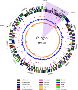
Rickettsia typi genome map. Credit: Virginia Bioinformatics Institute

Rickettsia typhi genome map. Credit: Virginia Bioinformatics Institute

ResearchBlogging.org
Maybe I am slow on the uptake, but I never quite liked the term “post genomic”, and I used it very sparingly. (Yes, I do have that term in one of my better cited papers, smack in the first sentence of the abstract, but I never liked that).  Perhaps because of all the associated abuse and hype that vacated the term from any core meaning it may have originally held; or perhaps it was because I saw genomics as an ongoing endeavor that will be embedded in life science for a long time to come, with no obvious “post” planned.  I even view metagenomics — hailed by many as a completely new and exciting field — as  an extension to genomics, with no clear boundary separating the two disciplines. So for me, a bioinformatician, genomics started somewhere around the mid 1990s when whole genome sequences started coming out, it is ongoing, and will continue, no “post-” in site.

Until now.

Continue reading My own post genomic moment →

Share and Enjoy:
  • Fark
  • Digg
  • Technorati
  • del.icio.us
  • StumbleUpon
  • Facebook
  • Reddit
  • Twitter
  • FriendFeed
  • PDF
  • email
  • Print
  • Google Bookmarks

Peace on Earth

Credit: Wikimedia Commons; image in Public Domain.

Here is a little trick I performed as a first year grad student a while ago and which I would like to share. My PhD adviser, Hanah Margalit was supposed to give a talk at a  joint Israeli-Palestinian meeting. (Hopefully, such days will be back soon and permanently!) The audience was rather broad, and included many non-scientists. So how do we show how cool bioinformatics  is to such a heterogeneous  crowd, in a space of 30 minutes, without losing their attention or going over their heads? Ron Unger from Bar Ilan university suggested we do what most of us always try to do: search for peace. Or rather, PEACE:  Proline, Glutamate, Alanine, Cysteine, Glutamate, a representation of a short peptide in one-letter amino acid code.

Continue reading Peace on Earth →

Share and Enjoy:
  • Fark
  • Digg
  • Technorati
  • del.icio.us
  • StumbleUpon
  • Facebook
  • Reddit
  • Twitter
  • FriendFeed
  • PDF
  • email
  • Print
  • Google Bookmarks

I’ve been slashdotted!

Slashdot article: Every man is an island (of Bacteria)

Share and Enjoy:
  • Fark
  • Digg
  • Technorati
  • del.icio.us
  • StumbleUpon
  • Facebook
  • Reddit
  • Twitter
  • FriendFeed
  • PDF
  • email
  • Print
  • Google Bookmarks

Genetic Algorithms

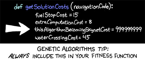
XKCD appeals to my geeky humor side. Also, as a staunch Pythoneer, I cherish the occasional Python reference. To complete the trifecta, I just saw T2 for the umptieth time two days ago.

Share and Enjoy:
  • Fark
  • Digg
  • Technorati
  • del.icio.us
  • StumbleUpon
  • Facebook
  • Reddit
  • Twitter
  • FriendFeed
  • PDF
  • email
  • Print
  • Google Bookmarks

Every Man an Island, Pt. 2

(Continued from  part 1)

ResearchBlogging.org

Why we are islands

In the previous post we have seen how  our bacterial population affects  our weight  and that by changing our dietary habits we can change the species composition in our guts. Also, we saw how a metagenomic analysis can lead to verifiable hypotheses: using a metagenomic analysis, Gordon’s lab discovered that the microbiome in the guts of obese mice have a high level of bacteria from the Firmicutes division; they also found that they contain a high level of carbohydrate-active enzymes or CAzymes.  These CAzymes break down sugars in our foods more efficiently, extracting more calories that contributes to weight gain in a vicious cycle.

Continue reading Every Man an Island, Pt. 2 →

Share and Enjoy:
  • Fark
  • Digg
  • Technorati
  • del.icio.us
  • StumbleUpon
  • Facebook
  • Reddit
  • Twitter
  • FriendFeed
  • PDF
  • email
  • Print
  • Google Bookmarks

Every Man an Island, Pt. 1

ResearchBlogging.org

No man is an island, entire of itself

— John Donne, Meditation XVII

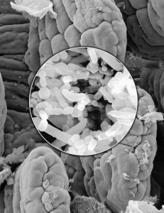
Scanning electron microscope images of <i>B. thetaiotaomicron</i>, a prominent human gut bacterium, and the intestine.  From: Human Gut Hosts a Dynamically Evolving Microbial Ecosystem Gross L PLoS Biology Vol. 5, No. 7, e199 doi:10.1371/journal.pbio.0050199
Scanning electron microscope images of B. thetaiotaomicron, a prominent human gut bacterium, and the intestine. From: Human Gut Hosts a Dynamically Evolving Microbial Ecosystem Gross L PLoS Biology Vol. 5, No. 7, e199 doi:10.1371/journal.pbio.0050199

Only one out of ten cells in our body is human

In a certain sense, every man is an island; this interesting finding comes from Jeffrey Gordon’s lab in Washington University. To understand why that is so, we need to understand something about the make up of our bodies. Adult human bodies are comprised of 1013 cells. These cells are broadly divided into different types that compose the tissues and organs that make us function the way we do. However, those are not all the cells that are in the human body. In addition to our own cells, we have 1014 bacterial cells that reside in and on us. Think about it: only one out of ten cells in our bodies contain the DNA inherited from our parents. The other nine cells are not human.

Continue reading Every Man an Island, Pt. 1 →

Share and Enjoy:
  • Fark
  • Digg
  • Technorati
  • del.icio.us
  • StumbleUpon
  • Facebook
  • Reddit
  • Twitter
  • FriendFeed
  • PDF
  • email
  • Print
  • Google Bookmarks

Blog for Darwin

I just went to Shirley Wu’s blog and found this. Apparently February 12-15, 2009 there will be a Blog for Darwin blogswarm, in honor of his 200th birthday and 150th anniversary of the publication of On the Origin of Species (First time I hear this term, blogswarm; not sure if I like it). I wrote about Darwin’s birthday earlier, but I guess I will write something new: my blog must adapt! (Actually, that is a very non-Darwinistic statement. If you figured out why, great. If not, answers will be provided sometime between Feb 12 and 15). Click on the image if you want to join the swarm.

Share and Enjoy:
  • Fark
  • Digg
  • Technorati
  • del.icio.us
  • StumbleUpon
  • Facebook
  • Reddit
  • Twitter
  • FriendFeed
  • PDF
  • email
  • Print
  • Google Bookmarks

Vaults

ResearchBlogging.org

They are very, VERY large (for intracellular particles, that is), there are hundreds of them most animal’s cells, and we don’t know what they are doing. Meet the vaults.

Vaults are mysterious large ribonucleoprotein (protein and RNA) structures in the cell. Although three times larger than ribosomes, and present in many copies in the cell, they were only discovered in the 1980s. Since their discovery some 25 years ago their cellular function, or functions, are still unclear.

In sea urchins, they have been shown to migrate to the nucleus. Because they are hollow and may fit well in the nuclear pore, some think they may be involved in nuclear transport. They have also been suspected to be associated with Multi Drug Resistance. MDR is an important phenomenon that has to do with sensitivity of tumor cells to cancer drugs. Tumors exhibiting MDR are less responsive to chemotherapy: they break down or remove cytostatic drugs. Vaults have been suspected of being such “drug shuttles” as they seem to be overexpressed in certain cancer cell types or when exposed to cytostatic drugs: drugs that are used to stop cells from dividing, common in cancer chemotherapy. Nevertheless, knockout mice studies have shown that mice where the gene from MVP is knocked are not extra sensitive to cytostatic drugs. However recently it was shown that vault particles may have a role in a related  cellular pathway,  programmed cell death, which may explain why they proliferate in cancer cell line, but do not affect sensitivity to cytostatic drugs that do not induce programmed cell death.

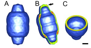
Why “vaults”?  Because when viewed through a longitudinal slice, they resemble a cathedral’s vaulted ceiling (See B, below).

Vault, Cryo-EM

Vault, Cryo-EM micrograph at 33A resolution. From Stewart et al. BMC Developmental Biology 2005 5:3 doi:10.1186/1471-213X-5-3. Clicking on link will take you to the original image in the article. Reproduced under the Creative Commons Attribution License

I am writing about vaults today because a group in Osaka, Japan has recently solved the structure of the vault in 3.5A resolution using X-ray crystallography. This still does not tell us what vault riboproteins do, but it may bring us closer now that we have a solution in atomic resolution. Also we get some really cool images and movies. The full PDB files can be downloaded form their site.  The main “construction unit” of the vault is the Major Vault Protein (MVP). The vault particle is composed from 78 MVPs  laid together as shown in the movie from the same site:

Here is a picture that shows a half vault, each of the 78 MVP chains colored differently, in spectral colors. One is drawn in white, for emphasis; looks a bit like like a Rasta wool cap, really.

vault-chaincolor1

Click for full size image. I colored one chain specifically in white, to show how it slants around the vault structure. Drawn using PyMOL

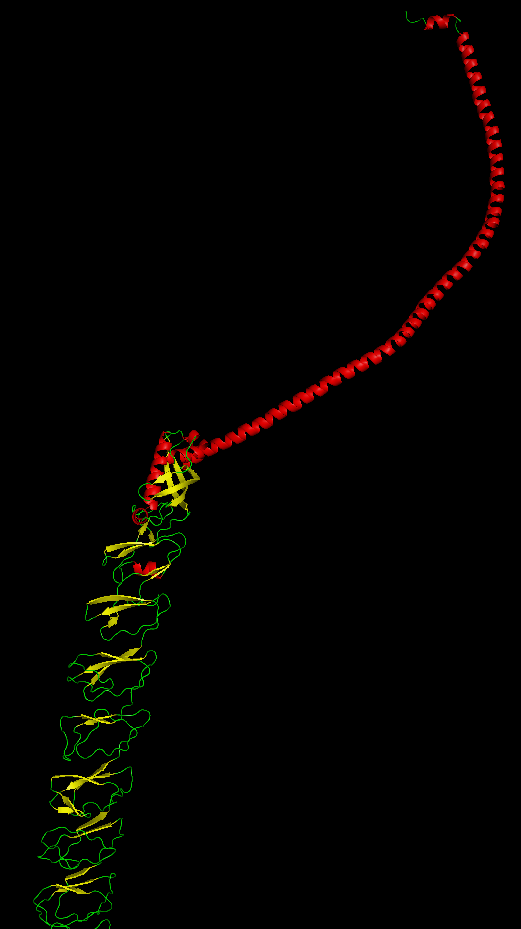
Last pic is of a single MVP chain, colored by secondary structures.

A single MVP.

A single MVP, notice how the repetitive stand (yellow) and loop (green) domains twist in relation to each other. Click for larger picture. Drawn using PyMOL.

So what are we left with? Literally, a big mystery. Very large cellular particles, almost everywhere; they do something, but after 25 years of knowing about them, we’re not sure exactly what. The Rome lab at UCLA has a very informative site about vaults. Happy vaulting.


H. Tanaka, K. Kato, E. Yamashita, T. Sumizawa, Y. Zhou, M. Yao, K. Iwasaki, M. Yoshimura, T. Tsukihara (2009). The Structure of Rat Liver Vault at 3.5 Angstrom Resolution Science, 323 (5912), 384-388 DOI: 10.1126/science.1164975

Share and Enjoy:
  • Fark
  • Digg
  • Technorati
  • del.icio.us
  • StumbleUpon
  • Facebook
  • Reddit
  • Twitter
  • FriendFeed
  • PDF
  • email
  • Print
  • Google Bookmarks